
Scientific Training I
Biomechanical Modelling. From Image Segmentation and Registration to Computational Meshing
PhD course covering image segmentation, registration, computational meshing, data analysis and how to obtain global accuracy in surface scanning of teeth.
| Place: | Faculty of Science, Frederiksberg Campus |
| Date and time: | 7 - 11 January 2019 |
| Seats: | 30 |
| ECTS credits: | 3 ECTS conditional on:
|
| Contact person / course organizer: | Sune Darkner, darkner@di.ku.dk |
| Language (written and teaching language): | English |
|
Course workload:
|
- Preparation: 50 hrs - Participation lectures: 10 hrs - Participation exercises: 10 hrs - Poster session: 5 hrs Total 75 hrs |
Program
Course material
See Internal Forum > courses > material t1 (section accessed only by project members)
Poster session
As part of the course program, a poster session is planned. If you wish to present a poster during the session, please submit a digital version on 7 January 2019 at the latest (to darkner@di.ku.dk) and bring a printed version for the course. Poster and presentation must reflect on how course content relates to own research. Poster and presentation must be approved by organizers to receive ECTS credit.
Course content
The scope of this course concerns the process of getting from a medical imaging to a computational model, a so-called mesh. The course is a mix of presentations of the latest theoretical cutting-edge developments in registration, segmentation and meshing combined with practical hands-on exercises on these topics. Through experience from the course and reflection in discussions with lectures and teaching assistants, the students will obtain practical working skills to apply in their own research projects.
The intended audience is PhD students with a computer science or engineering background or similar. Programming in matlab, python or C++ may be part of the exercises, and lectures may contain mathematical high level content. Master students are welcome as well, particularly if their master thesis projects are aligned with this course. Audience with background in physics and mathematics or similar fields may find this course useful as well, but rudimentary programming skills are necessary to complete some of the exercises.
Generally, medical diagnosis, prognosis and quantification of progression are based on biomarkers. These may be blood or urine markers, but currently imaging is taking over as a more indicative biomarker for many purposes. This course will offer an introduction to medical image formation in the different scanning modalities. We will continue with the underlying image analysis disciplines of detection, registration, and segmentation, and end with specific applications in clinical practise. A key to achieve success in the medical image analysis is formal evaluation of methodologies, thus an introduction to performance characterisation will also be a central topic. We will use techniques from image analysis and real world examples from the clinic, with particular outset in the research projects in RAINBOW MSCA-ITN.
Learning outcome
Knowledge
- Image Segmentation
- Image Registration, for instance non-rigid registration based on deformable models
- Computational Meshing: Mesh and polygonal mesh generation, contouring and quality mesh approaches using deformable simplicial complexes.
Skills
- Applying methods from image segmentation, registration, shape modelling and computational meshing to acquire high quality computational meshes from image data.
Competences
- Build systems for rapid biomechanics simulation for personalized clinical design,
- Apply such software systems for solving problems relevant to industrial health business cases.
Program
| Time | Monday 7 Jan. |
Tuesday 8 Jan. |
Wednesday 9 Jan. |
Thursday 10 Jan. |
Friday 11 Jan. |
| A1-34.22 | A2-71.02 | A2-71.01 | A1-37.11 | A2-71.02 | |
| 9:00 | Sune Darkner: Registration. 35 min. | Francois Lauze: General introduction to segmentation, Mean shift, K-means clustering etc.. Good continuation and Snakes. 35 min. | Vedrana A. Dahl (VAD): introduction to segmentation: Shape models vs pixel level segmentation. 35 min. | Peter Søndergaard and Peter Dahl: Obtaining global accuracy in surface scanning. 35 min. | Sune Darkner: Machine learning. 35 min. |
| 9.35 - 9.50 Break |
9.35 - 9.50 Break |
9.35 - 9.50 Break |
9.35 - 9.50 Break |
9.35 - 9.50 Break |
|
| 10:00 | Registration continued. 35 min | Contour based methods, Balloon forces, Geodesic Active Contours, Levelset method. 35 min. | Andreas Bærentzen (AB): Scalar fields and distance fields. Methods and applications. Simplicial complexes. Definitions and properties. 35 min. | Obtaining global accuracy in surface scanning (continued) 35 min. | Machine learning continued. 35 min. |
| 10.25 - 10.55 Break |
10.25 - 10.55 Break |
10.25 - 10.55 Break |
10.25 - 10.55 Break |
10.25 - 10.55 Break |
|
| 11:00 | Registration continued. 45 min | Region based method. The Mumford-Shah functional, The Chan and Vese approach. Levelset formulation of the Chan and Vese, as well as Regularized labelling formulation. 45 min. | VAD: The Mumford-Shah functional and methods. Dictionary based segmentation. 45 min. | Obtaining global accuracy in surface scanning (continued) 45 min. | Machine learning continued. 45 min. |
| 11.40-11.55 Break |
11.40-11.55 Break |
11.40-11.55 Break |
11.40-11.55 Break |
11.40-11.55 Break |
|
| 12:00 | Registration continued. 45 min. | Faezeh Moshfeghifar and Susanne Claus: Presentation of case study of hip joint modeling. 45 min. |
AB: Introduction to topologically adaptive deformable interface tracking: |
Obtaining global accuracy in surface scanning (continued). 45 min. | Machine learning continued. 45 min. |
| 13:00 | 12.40 - 13.30 Lunch |
12.40 - 13.30 Lunch |
12.40 - 13.30 Lunch |
12.40 - 13.30 Lunch |
12.40 - 13.30 Lunch |
| 14:00 | José Tascon: presentation of Kitware case. 2 hrs. | Exercise 2 hrs. | Exercise: 2 hrs. Supervised by TAs: - practical dictionary based segmentation - practical DSC based segmentation. - no coding - just experiments with image data sets and parameters |
Exercise 2 hrs. |
Exercise 1½ hr. 14:00 - 15:00 Christian Wong: 'Finite element simulation to clinical application' |
| 15:00 | 15:00-15:30 Break |
15:00-15:30 Break |
15:00-15:30 Break |
15:00-15:30 Break |
15:00 Course closure |
| 16:00 | 15:30-16:30 Kitware case continued |
15:30-16:30 Exercise continued |
15:30-16:30 Exercise continued |
15:30-16:30 Exercise continued |
|
| 17:00 | Poster Session 17:00-19:00 |
Walk and pre-dinner beer | |||
| 18:00 | Dinner 18:30 |
Invited Guest Lecturer 1:
Andreas Bærentzen
 Andreas Bærentzen is an associate professor in computer graphics at the Department of Applied Mathematics and Computer Science at DTU. He received his MSc Eng and PhD degrees from the Technical University of Denmark in 1998 and 2003 respectively. Andreas' research interests are in computer graphics, and in particular modelling and manipulation of shapes and real time graphics. He is especially interested in representing shapes in ways that allow for topological adaptivity, interactive modeling, and efficient geometry processing.
Invited Guest Lecturer 3:
Peter Dahl

Peter Dahl is a Software Developer at the Trios Software team in 3Shape Trios A/S, where he develop a scanning application that receives data from the intraoral scanner Trios and reconstructs it to a 3D scan. Peter holds a M.Sc. in applied mathematics from DTU and has been with 3Shape for 9 years. During his time in the company he has worked on nearly every part of the reconstruction engine, but primarily focused on the real-time scanning feedback, GPU processing and general application development. He has also been involved in a project with the Audio team where the same scanning technology were adapted to scan ears instead of teeth. Before coming to 3Shape he worked in the computer games industry for a few years as a graphics programmer.
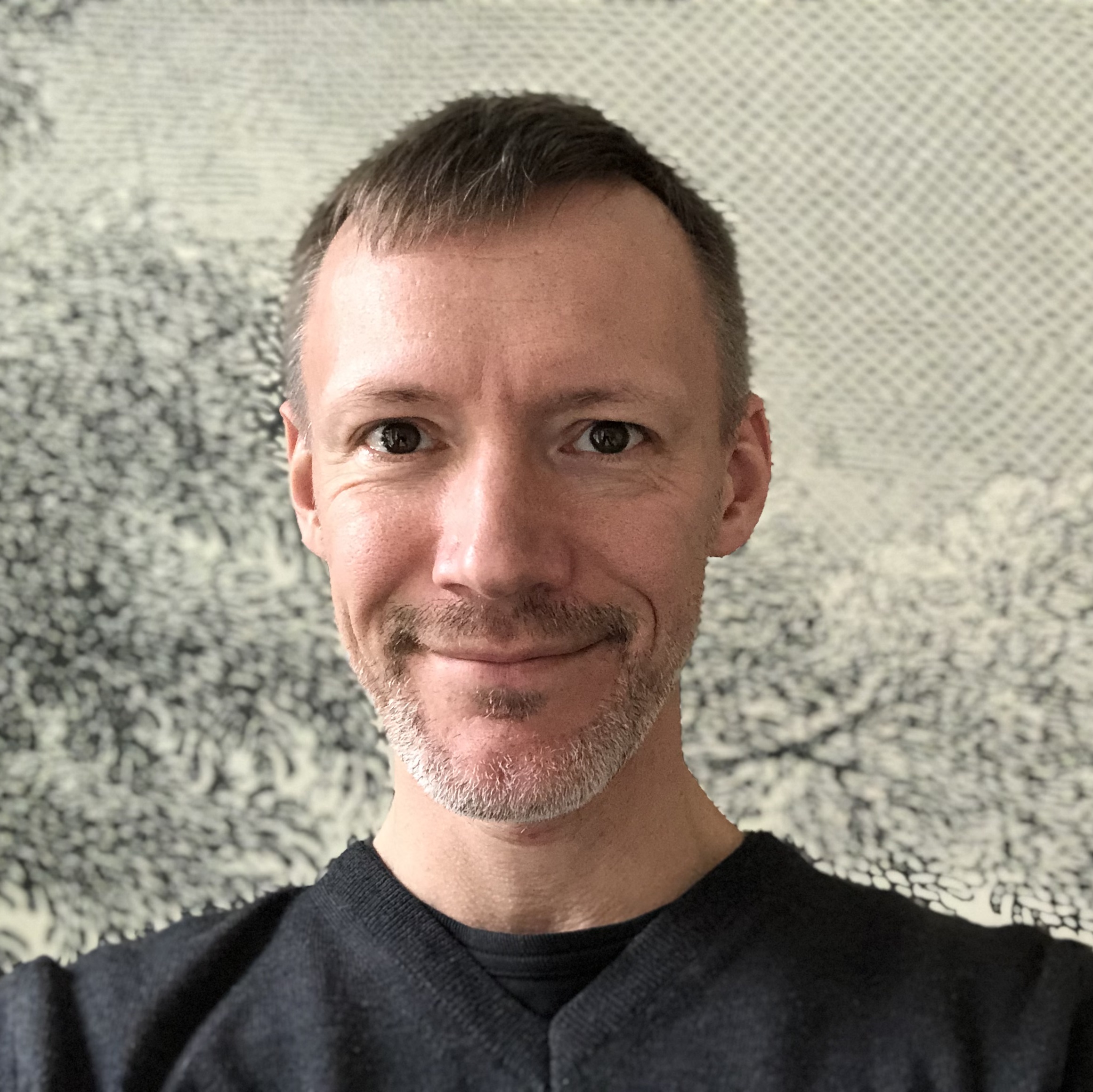
Invited Guest Lecturer 5:
Sune Darkner
 Sune Darkner is associate professor at KU since 2012. His main research topic is medical image. He work mainly on neuro-imaging data such as MRI and PET and holds expertise on image registration and the statistics on shapes and deformations in relation to simulation of soft tissue. He has published more than 40 journal and conference papers, has co-organized workshops under MICCAI and is co-supervising 1 PhD, 2 others finished primo 2015. He is member of RAINBOW's Supervisor Board and supervisor of ESR 2 and ESR 4.
Further profile information on Sune Darkner.
Invited Guest Lecturer 2:
Vedrana Andersen Dahl
 Vedrana Andersen Dahl is an associate professor at the Department of Applied Mathematics and Computer Science, Technical University of Denmark. She holds degrees in mathematics, multimedia technology, and a Ph.D. degree in geometry processing. Her research interests revolve around geometric models for analysis of volumetric data. This includes volumetric segmentation, tomographic segmentation and methods based on deformable meshes. She developed image analysis tools with application in material science, industrial inspection and biomedicine. Her other research interest involve 2D and 3D texture analysis and 3D methods for digitizing cultural heritage. Further profile information.
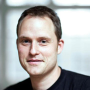
Invited Guest Lecturer 4:
Peter Søndergaard
 Peter Søndergaard is team lead for the Orthodontics Technology Team at 3Shape A/S, specializing algorithmic and machine learning solutions to orthodontic problems to be medically validated.
Peter has a M.Sc. and ph.d. from DTU in mathematics, specifically time-frequency analysis and wavelet theory. He has worked 5 years as a post.doc, assoc. prof. and group leader at the Centre for Applied Hearing Research (DTU, Lyngby) and Austrian Academy of Sciences (Vienna, Austria) investigating fundamental problems in the mathematical modelling of human hearing and 4 years at Oticon A/S as audiological specialist and project manager. He is member of RAINBOW's Supervisor Board and supervisor of ESR 3. Further profile information on Peter Søndergaard.
Invited Guest Lecturer 6:
Francois Lauze
 Francois Lauze is associate professor at Department of Computer Science where he specializes in Mathematical Image Analysis, including variational and partial differential equations methods, with applications to image inpainting (still and video), motion estimation in image sequences, segmentation and video format conversion. Further, he studies differential and Riemannian geometric methods for shape analysis applied to anatomical structures extracted from X-ray and other medical imaging modalities. Further profile information on Francois Faure.
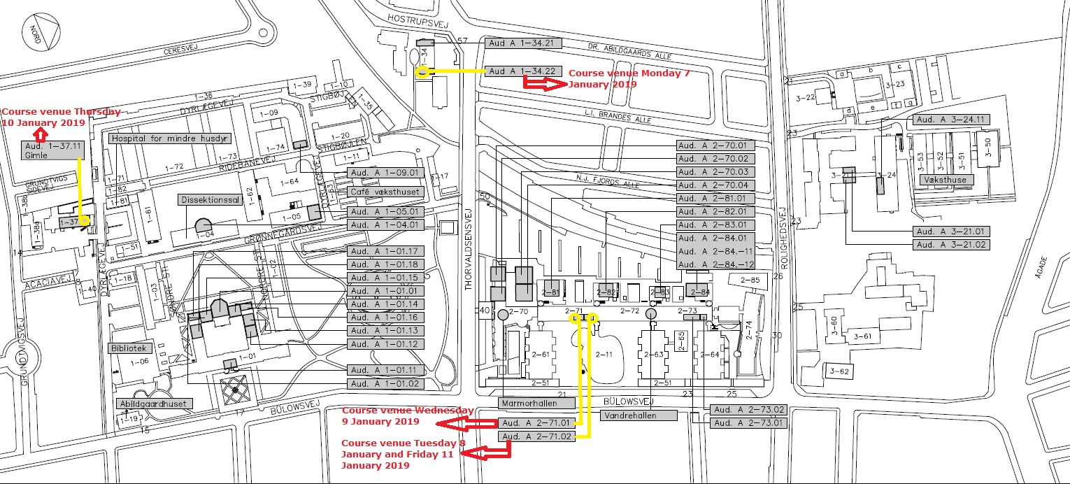
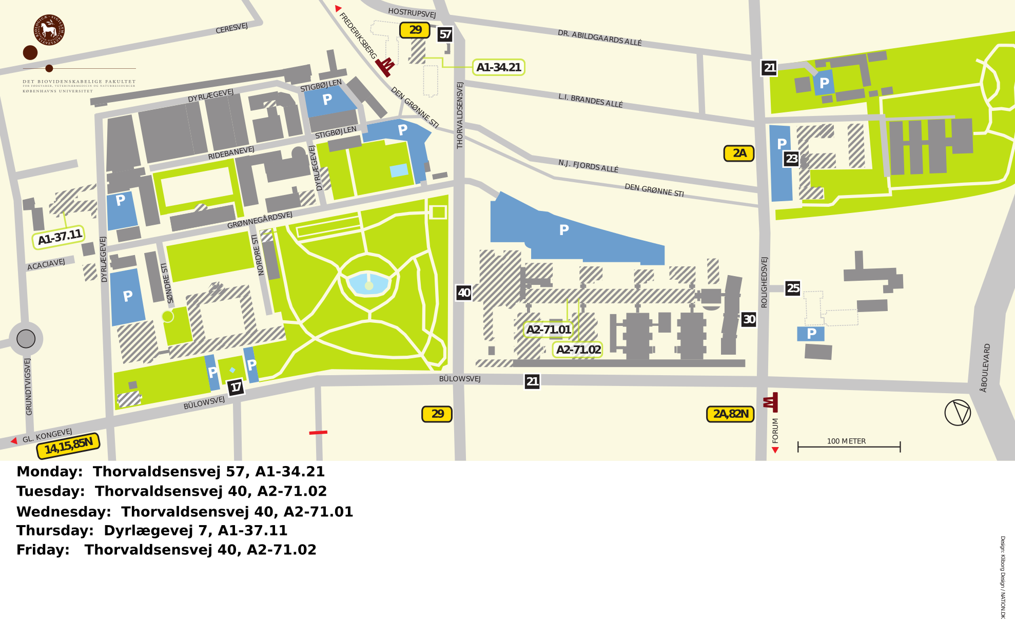
Course dates and venue
7 - 11 January 2019
Venue: Frederiksberg campus
See map of campus.
Rooms for each day listed in the program.
Registration
Registration deadline: 20 December 2018
Note: Travel and accommodation to be booked individually by participants.
Reasonably priced hotels are found in this list.
Practical information
- University of Copenhagen's locations
- public transport
- hotels
- tourist information